
3D IMAGING AND DIGITAL DIAGNOSTICS (X-rays, CT)
3D IMAGING AND DIGITAL DIAGNOSTICS (X-rays, CT)
Modern dentistry relies heavily on advanced imaging technologies. Accurate diagnostics are essential for planning safe, precise and predictable treatments. Our clinic is equipped with state-of-the-art diagnostic systems, allowing us to provide comprehensive examinations and create highly detailed, personalised treatment plans.
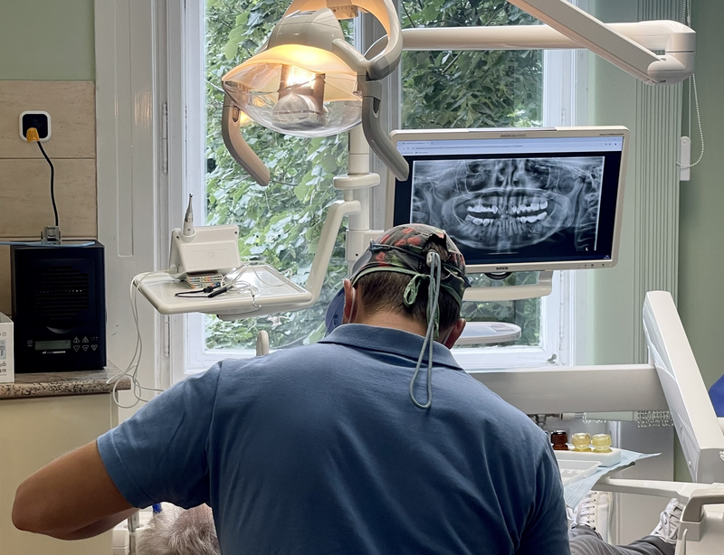
DENTAL X-RAYS
Dental X-rays are an indispensable part of everyday dental diagnostics, as they allow us to examine the teeth, roots, jawbones and surrounding structures in detail, often revealing conditions that are not visible during a regular clinical examination. Depending on the purpose of the examination, we may use intraoral or extraoral X-rays, each providing different types of information for diagnosis and treatment planning.
There are two main types of dental X-rays: intraoral and extraoral images.
Intraoral X-rays are taken inside the mouth and focus on a specific tooth or group of teeth. They are used to detect cavities, examine the roots and root canals, and identify inflammation or other changes around the teeth.
Extraoral X-rays provide a broader overview of the oral and maxillofacial region. A panoramic X-ray is often used as an initial diagnostic image, while a lateral cephalometric X-ray is commonly used in orthodontics to evaluate the relationship between the upper and lower jaws and the skull. It can also play an important role in planning the surgical correction of bite irregularities.
CBCT
CBCT, or cone beam computed tomography, is a modern 3D imaging technique that has significantly improved dental and oral surgical diagnostics. It provides highly detailed, three-dimensional images of the teeth, jawbones and surrounding anatomical structures.
This allows us to plan complex procedures with a high level of precision, including wisdom tooth removal, implant placement and advanced bone grafting procedures.
How does 3D imaging elevate your treatment?
- Precise diagnostics: 3D imaging helps our dentists and oral surgeons establish diagnoses more quickly, accurately and confidently.
- More predictable treatment planning: It supports the assessment of wisdom teeth and supernumerary teeth, the evaluation of bone volume before implant placement, the planning of bone grafting procedures, and the detection of complex root canal anatomy.
- Clear, distortion-free visualisation: Anatomical structures can be viewed from multiple angles without distortion or overlapping images.
- Better patient communication: 3D images make diagnoses and treatment plans easier to explain and understand, helping patients make informed decisions.
Beyond that, 3D imaging is also invaluable in implant dentistry: implants can be planned virtually before surgery, allowing for optimal positioning and safer, more predictable outcomes.
HOW CAN 3D INTRAORAL SCANNING MAKE TREATMENT MORE PRECISE AND COMFORTABLE?
The intraoral scanner is an innovative digital device that allows us to take fast, painless and highly accurate digital impressions. Using a small handheld scanner, we create a detailed 3D image of the teeth and oral cavity, providing a precise digital model for treatment planning.
This technology is not only more comfortable for patients, but also enables the creation of highly accurate restorations, such as crowns, bridges, veneers and implant-supported prosthetics, as well as clear aligners. Intraoral scanning is especially useful in orthodontics, aesthetic dentistry and implant treatments, where precision and digital planning play an important role.
At our clinic, we use advanced digital dentistry solutions to make treatments more comfortable, predictable and efficient, while ensuring that each patient receives care tailored to their individual needs.