
Imaging Diagnostics (X-rays, CT)
Imaging procedures are essential for establishing the correct diagnosis and treatment plan, allowing us to provide comprehensive examinations for our patients.
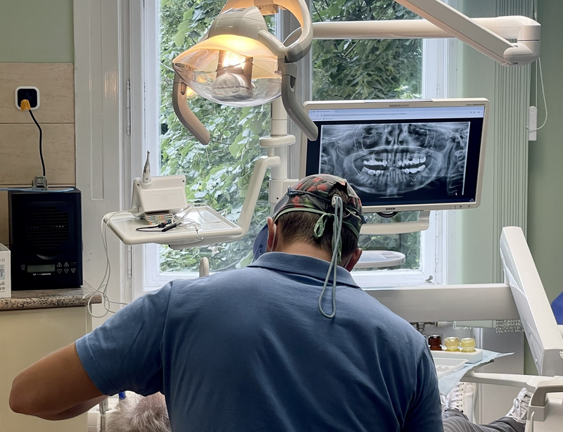
X-rays:
We differentiate between extraoral and intraoral X-rays, meaning those taken outside and inside the mouth, respectively.
During intraoral X-ray imaging, a targeted image of a specific tooth or group of teeth is taken to visualize conditions such as cavities, roots, root canals, and inflammation around the teeth.
Extraoral X-rays provide a more comprehensive view of the oral cavity. A common type is the panoramic X-ray, often used as an initial diagnostic tool, and the lateral X-ray, frequently employed in orthodontics.
CT
3D imaging offers several advantages:
- Precise diagnostics: Our dentists and oral surgeons can diagnose conditions more easily, quickly, and confidently.
- Assistance with diagnosing supernumerary teeth, impacted wisdom teeth, determining bone availability for implant placement, planning bone grafting procedures, and locating difficult-to-find canals during root canal treatment.
- Lifelike visualization: Anatomical structures can be visualized from any angle without distortion or overlap.
- 3D visualization aids communication with patients, making diagnoses easier to understand.
The usability of 3D imaging systems in orthodontics includes:
- 3D examination of tooth positioning, precise visualization of impacted wisdom teeth and supernumerary teeth.
- Analysis of tooth and jaw growth.
- Planning for orthodontic implant placement.
- Scanning of plaster models before creating digital models.
- Designing custom orthodontic appliances.
Implant planning:
- Implants can be placed in the best possible positions for the intended prosthetic restoration.
- Virtual implant placement before surgery helps with planning.
- Possibility of creating 3D-printed surgical guides.
Dental 3D imaging systems contribute to accurate diagnosis and efficient treatment planning. Patients should attend regular check-ups and follow the doctor’s instructions for treatment or maintaining oral health. These modern imaging systems enable doctors to assist patients more effectively and safely in preserving their healthy oral cavities.